The retina is the innermost nerve layer that lines the back of the eye.
Many ophthalmologists compare this to the film of a camera. The retina is responsible for processing the images projected onto it and then the optic nerve transmits this to the brain. The retina is susceptible to many
types of diseases.
What are the common problems associated with the retina?
Retinal detachment
A retinal detachment is a very serious problem that almost always causes blindness unless it is treated.
As a person’s normal eye ages the vitreous gel contracts and then becomes more liquid.
As the vitreous gel becomes more liquid it may pull on the retina and create a retinal tear (and then tear.)
When fluid passes through a tear, it will lift the retina from the back of the eye creating (making) a retinal detachment.
Macular Degeneration
Macular Degeneration, also called age related macular degeneration (ARMD), affects primarily the aging population and gradually destroys central vision.
As noted above the central vision is critical for seeing fine details and necessary for performing daily tasks such as reading and driving.
This degenerative eye disease causes no pain and typically develops over a long period of time.
Patients may notice a blurring or distortion in their central vision.
In other words, lines that should be straight may appear to have a bend in them.
Diabetic Retinopathy
Diabetic retinopathy is a leading cause of blindness.
In the majority of diabetic retinopathy cases, blindness is completely preventable.
The use of medications and daily blood sugar monitoring can make a major impact on preventing or containing any worsening of diabetic retinopathy.
Flashes/Floaters
Floaters may appear as small specks or dark shadows.
These specks can actually move or float around in the visual field.
Most people have some level of floaters and normally do not notice them until they become very obvious.
Sometimes floaters can be seen when you stare up at the sky and they may appear as curvy lines or small specks.
This condition actually develops from changes in the back of the eye also known as the vitreous cavity.
In typical cases floaters are part of the aging process.
Macular Edema
Macular Edema is also called Cystoid Macular Edema or (CME). Macular Edema sets in when blood vessels leak into the eye and form cysts and swelling that obstruct central vision. CME can best be seen from a diagnostic vision
test called fluorecein angiography or optical coherence tomography.
Retinitis Pigmentosa
Retinitis Pigmentosa refers to a group of inherited eye diseases causing the degradation of the retina.
As we have discussed throughout the retina section of this website, the retina is a critical component of the eye responsible for interpreting images and transmitting them onto the brain.
When retinitis pigmentosa develops, the cells, which are called rods and cones, die.
The loss of visual fields is the indicator for retina pigmentosa and begins as a ring like distortion in the mid-periphery.
Throughout the course of this degenerative eye disease the central vision is also lost and the result is tunnel vision.
As a result of this progression patients are told not to drive automobiles even in the early stages.
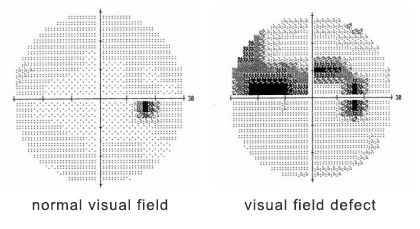
Perimetry
Perimetry is the systematic measurement of visual field function.
The two most commonly used types of perimetry are Goldmann kinetic perimetry and threshold static automated perimetry.
With Goldmann or "kinetic" perimetry, a trained perimetrist moves the stimulus; stimulus brightness is held constant.
The limits of the visual field are mapped to lights of different sizes and brightness.
With threshold static automated perimetry, a computer program is selected.
The most commonly used one tests the central 30° of the visual field using a six degree spaced grid.
This is accomplished by keeping the size and location of a target constant and varying the brightness until the dimmest target the patient can see at each of the test locations is found.
These maps of visual sensitivity, made by either of these methods, are very important in diagnosing diseases of the visual system.
Funduscopy
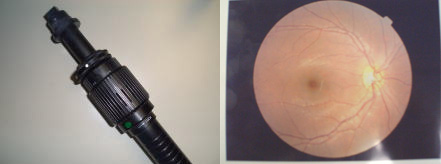
Also termed ophthalmoscopy. The examination of the back of the eye known as the fundus.
The examination is carried out to check eye health and to ensure that there are no signs of retinal detachment,
retinal holes or other eye problems such as diabetic retinopathy and glaucoma.
The examination may begin with dilating the pupil so that examination is easier.
The back of the eye is illuminated using a beam of light through a funduscope.
Fluorescein fundus photography
This is a technique for examining the circulation of the retina using the dye tracing method.
It involves injection of sodium fluorescein into the systemic circulation, and then an angiogram is obtained by photographing the fluorescence emitted after illumination of the retina with blue light at a wavelength of 490 nanometers.
The fluorescein dye also reappears 12-24 hours in the patient urine, causing a yellow-green appearance.
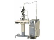
OCT (Optical Coherence Tomography)
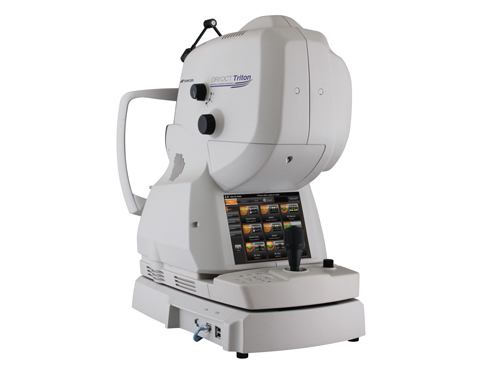
OCT (Optical Coherence Tomography) is a test to shoot a fault screen of the retina.
This test clarifies the condition of the retina, which is difficult to diagnosis, and helps to decide treatment plans for retina disease.
OCT is necessary for diseases such as macular hole, diabetic maculopathy, macular edema, age-related macular degeneration, detachment of the retina, and glaucoma.
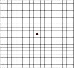
Amsler Grid
The Amsler grid is a tool that eye doctors use to detect vision problems resulting from damage to the macula (the central part of the retina) or the optic nerve.
The damage may be caused by macular degeneration, glaucoma or other eye diseases, so the Amsler grid is useful in detecting these problems.
An early diagnosis means early treatment, so it may help to limit or at least slow the vision loss you experience.
If you are at risk for macular degeneration or other eye diseases, you can use this chart at home to monitor your vision.
But using the chart doesn’t mean you should skip regular visits to your eye doctor, because you can easily miss signs that only a trained eye care practitioner will find.
The chart below is an approximation of the printed chart used by eye doctors.
For more accurate detection of macular damage, you will need a complete eye exam.
Treatment of Retina
If you have retina detachment or retinal break, you need to operate your eye.
| State of the retina detachment
| type of operation
| contents of operation
|
| become split your retina |
Photocoagulation |
close up the surrounding your eye crack
with the laser light |
enter into liquefied vitreous body
and peel off your retina |
Retinopexy |
put your peeled retina back in its place |
bleed to vitreous body from blood vessel
when your retina will split |
Vitreous surgery |
remove your impure vitreous body by bleeding |