


Director:Yasuhiro Shinkawa
(Board-Certified Ophthalmologist by the Japanese Ophthalmological Society)
Memberships
Japan Ophthalmological SocietyJapanese Retina and Vitreous Society
Japanese Society of Ophthalmic Surgeons
Certification of Completion
Course of Ophthalmic PDT Study GroupNumber of cataract surgery up to the present:About 4000
Career
2001 Graduate-Medical Department of Kumamoto University2002 Department of Ophthalmology Kyoto University School of medicine
2002 Shimada Municipal Hospital
2008 Japanese Red Cross Society
2010 Kitano Hospital The Tazuke Kofukai Medical Research Institute
2014 Shinjuku-Higashiguchi Eye Clinic
Doctor:Fumiyo Hasegawa
(Board-Certified Ophthalmologist by the Japanese Ophthalmological Society)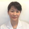
Memberships
Japan Ophthalmological SocietyJapan Ophthalmologists Associasion
Japanese Association for Strabismus and Amblyopia(JASA)
Career
1992 Graduate- Medical Department of Teikyo Univercity2002 The head ophthalmologist at International Catholic Hospital
2020 Shinjuku-Higashiguchi Eye Clinic
Main Thesis
Sequelae of ocular trauma in schools.(Japanese)A case of periodic upper and lower strabismus with loss of periodicity after cataract surgery(Japanese)
Quantitative analysis of eye movement during a cover test for patients with intermittent exotropia(Japanese)
Another several ophthalmologists are working here.