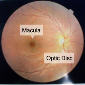
A macular hole is a small break in the macula, located in the center of the eye’s light-sensitive tissue called the retina. The macula provides the sharp, central vision we need for reading, driving, and seeing fine detail.
A macular hole can cause blurred and distorted central vision. Macular holes are related to aging and usually occur in people over age 60.
What causes a macular hole?
Most of the eye’s interior is filled with vitreous, a gel-like substance that fills about 80 percent of the eye and helps it maintain a round shape. The vitreous contains millions of fine fibers that are attached to the surface of the retina. As we age, the vitreous slowly shrinks and pulls away from the retinal surface. Natural fluids fill the area where the vitreous has contracted. This is normal.
If the vitreous is firmly attached to the retina when it pulls away, it can tear the retina and create a macular hole. Also, once the vitreous has pulled away from the surface of the retina, some of the fibers can remain on the retinal surface and can contract. This increases tension on the retina and can lead to a macular hole.
Macular holes can also occur in other eye disorders, such as high myopia (nearsightedness), injury to the eye, retinal detachment, and, rarely, macular pucker.
What are the symptoms of a macular hole?
In the early stage of a macular hole, people may notice a slight distortion or blurriness in their straight-ahead vision. Straight lines or objects can begin to look bent or wavy. Reading and performing other routine tasks with the affected eye become difficult.
Examination
Retinal Examination(Ophthalmoscopy)
This exam needs to open your pupil. Eye drops may be placed in your eyes to widen (dilate) your pupils. This test can help to determine cataract and another inflammation of crystalline lens.
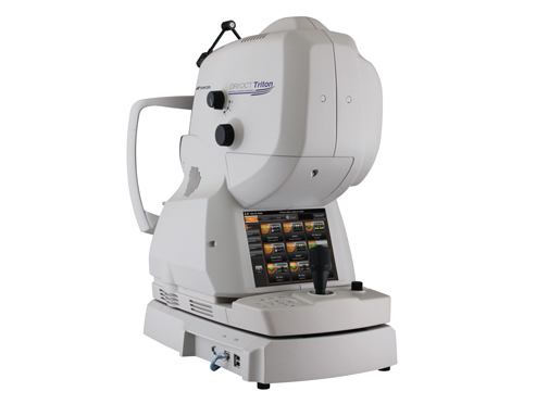
DRI OCT Triton
Take photograph of retina. This test can help to determine detailed data of your eyes like a MRI. This test is used to examine the glaucoma and loss of eyesight.
What’s the treatment for a macular hole?
Although some macular holes can seal themselves and require no treatment, surgery is necessary in many cases to help improve vision. In this surgical procedure – called a vitrectomy – the vitreous gel is removed to prevent it from pulling on the retina and replaced with a bubble containing a mixture of air and gas.
- 1.Make three small holes on the eye, and then remove the vitreous gel with a vitrectomy cutter.
- 2.Remove a thin membrane which is attached to around macula.
- 3.In the case that there is any tear on the other part except for the retina, have a laser coagulation.
- 4.Replace the vitreous gel with a bubble containing a mixture of air and gas. After surgery, the patient needs to keep lying faced down for a week to let the holes be closed by ballooning power of the gas. The gas will be absorbed naturally and be replaced with the aqueous humor secreting into the eye.