Contact Lenses
Contact Lens Troubles
Please refer the contents below if you are in urgent trouble.
What should we do when we can’t take off contact lens from eyes?
- Try to search contact lens
Please observe your eye carefully and check if the lens in your eye.
Contact lenses never go to back side of eyeballs. Try to search lens by moving your eyeballs slowly up and down, left and right with looking in a mirror.
When you can’t find lens in your eye, you may have dropped off lens. Even in that case, you still need to go to hospital to make sure there’s no problem.
- In the case lens sticks to eye
Please apply an eye drop if you have one. If you don’t have any, just close your eyes for a while and moisten your eyes with your tears.
When the lens places in the center of cornea, please try again to take off lens in usual way. In the case still you can’t take off, please hold down eyelid gently with your eyes closed and move your eyeball to the side of ear. And then, lens will come rising up.
If the lens does not come off even with those, please try to blink in the water filling washbowl.
- Please consult Shinjuku-Higashiguchi Eye Clinic
If you can’t take off lens with all effort, you need to see doctor. Please contact us when there remaining hyperemia or pain.
Things to prepare when you come to our clinic
- Health insurance card
- Contact lens data you use
- Eye drops usually you use
- Appointment will make your consultation smooth
Phone
03-5363-0507
+81-3-5363-0507
Online
When you miss contact lens in your eye
-
You have no idea if there remain contact lens or already come off.
At first don’t be confused, look into the mirror and confirm if the lens is there. When you can’t find, please move your eyeballs up and down, left and right to search on the white of eye or edge of the eyelid. Contact lenses never go to back side of eyeballs structurally. If you see clearly with one-eye hidden, there may remain the lens on your cornea.
-
You can’t find lens even though there is foreign body sensation
Look to opposite side which you feel foreign body sensation and search the contact lens. Try to blink in the washbowl filled with water. This procedure might improve your foreign bode sensation by washing away the pieces of contact lens. Please don’t try to remove contact lens forcibly. There sometimes causes scar on your eye if you scratch them.
Things to prepare when you come to our clinic
- Health insurance card
- Contact lens data you use
- Eye drops usually you use
- Appointment will make your consultation smooth
Phone
03-5363-0507
+81-3-5363-0507
Online
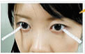
How to deal when lens cleaning solution get into your eye…
First aid
Rinse your eyes with a lot of water and see the ophthalmological doctor immediately bringing the package of solution.
Soft lens solution
Multi purposes solution don’t hurt your eye because concentration of disinfection ingredient is low.
On the other hands, soaking-disinfection type sometimes occur injury to your eyes if they come into eyes.
Most of these solution contains hydrogen peroxide as a disinfection ingredient. Hydrogen peroxide is an ingredient which is used as oxidizer or bleaching agent. It has strong effect to disinfection so it causes pain if it comes into your eye without neutralization. In worst case, it causes serious eye disease and even losing eyesight. So it is necessary to neutralize solution before wearing lens.
Hard lens solution
Many of solution for hard lens contain surfactant and enzyme. Surfactant is an ingredient which is used for detergent. They also can cause cornea disease if come into eyes.
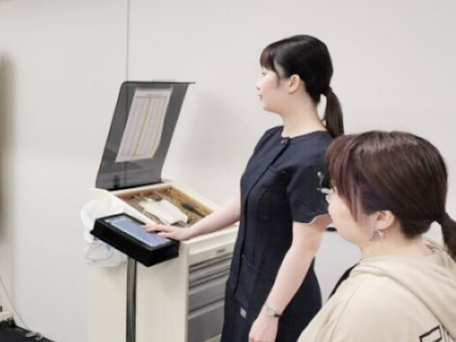
The flow for patients contact lens problems.
Flow from reception to payment
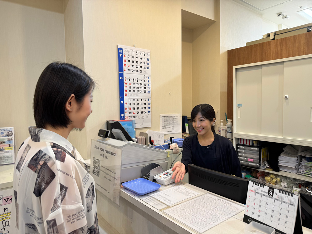
1.Reception
When you come to the clinic, please bring your insurance card if it is your first visit, or your consultation card if it is a follow-up visit, and present it to the receptionist. Please fill out the medical questionnaire, while we keep your insurance card. Please fill in all items on the medical questionnaire.

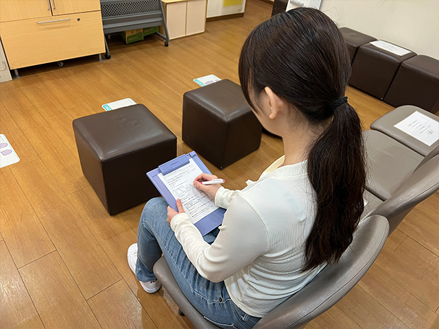
When you have filled out the medical questionnaire, the examiner will verbally confirm details before the basic examination, such as when and in which eye you have symptoms, whether you have any history or family history, including systemic diseases, what type of contacts you are using, and whether you are allergic to any medications.
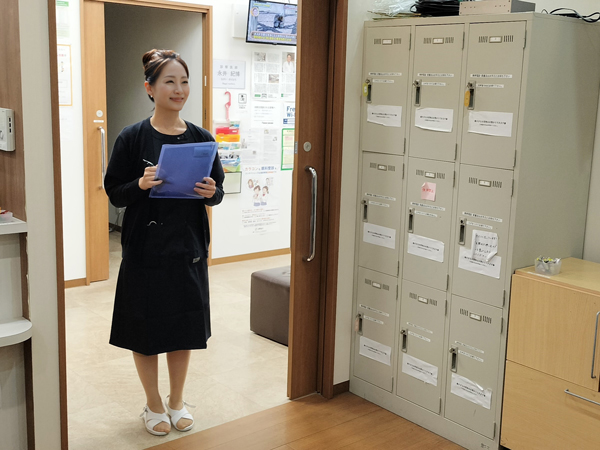
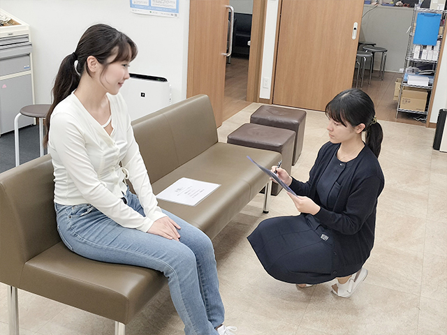
We will quide you to the examination room, and check your medical questionnaire.
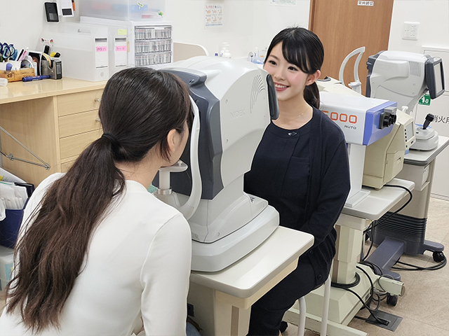
2.Examinations
We will exam your refraction, intraocular pressure, and your corrected vision.
-
-
What is a refraction test?
We wiil generally check how much hyperopia, myopia, astigmatism you have, and exam your corrected vision according to the number.
What is an intraocular pressure test?
Since some patients may experience an increase in intraocular pressure with eye drops, we will take data of your original intraocular pressure at the beginning of the examination. Based on these numbers, we will determine if it is safe to use eye drops or if other tests are necessary.
What is a corrected vision?
-
-
Based on the refraction test, we will examine whether your vision is good up to 1.2. This test is used to determine if there are any scars on your eyes which may be causing difficulty in achieving good vision, or if there are any hidden diseases other than contact lens trouble. It is different from the power of contact lenses or glasses, so if you would like a prescription, please come back after your symptoms have healed.
There are various examination and it may be performed as needed.
Schirmer’s test
People who wear contact lenses often complain of dry eyes. The Schirmer test measures the amount of tears produced.
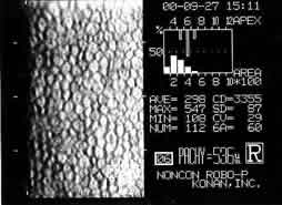
Specular Microscopy
It is said that long-term use of contact lenses reduces the number of endothelial cells in the innermost part of the cornea compared to those who do not use contact lenses. Using the specular microscopy, we will check if you have enough amount of endothelial cells.
※If you have any question about the examination, please feel free to ask the staff.
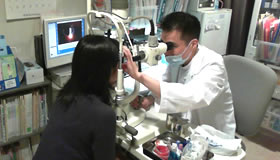
3.consultation
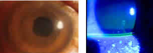
The doctor will examine the patient based on a medical interview and basic examination. In the consusltation room, fundus examination will be performed using a slit lamp microscopy.
Slic Lamp Microscopy
-
-
The patient will be asked to place the chin on the chin rest to examine whether the surface of the eye is damaged. In some cases, tears will be dyed to make it easier to see surface flaws.
Fundus Examination
The lens will be placed in front of the eye to examine the fundus. If there is any abnormality in the fundus, a more detailed examination may be performed.
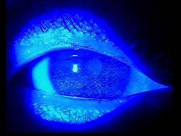
Diseases that could be found by consultation
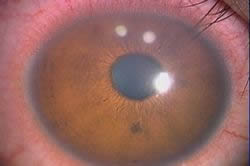
Corneal Epithelial Defect
Although there may be no symptoms in some cases, the patient may experience constant tearing, pain, redness, eye discharge, or part of cornea will become white and cloudy. Contact lense deposits, or care products can damage the epithelium, causing pain and redness.
superficial punctate keratitis
-
-
As the name suggests, it causes dot-like damage spot on the cornea. This eye disorder is a common disease caused by contact lenses.
Wearing contact lens for long time or a dirty lens would disturb an oxygen circulation on the cornea.
The disorder will recover soon after stopping wearing contact lenses. However, it also easily appears again unless the cause of syptoms is eliminated.
If it gets worse, a damage on the cornea would be deep. A corneal damage may cause more severe symptoms such as corneal epithelial erosion, corneal infiltration and corneal ulcer.
smile mark staining
-

-
People who wear contact lenses for long period of time often get a smile mark staining. Why it is called as “”smile mark”” is because the damaged area on a cornea seems like the shape of a smiley mouth.
The symptoms may be caused by wearing high oxygen permeable contact lenses, dry eye and incomplete blink. It would cause foreign body sensation in eyes, but it is easily ignored because people would feel less irritiation while wearing contact lenses. For complete treatment, patients will need to stop wearing unsanitary lenses, and reduce time waering contact lenses.
corneal endothelial disorder
-
-
The most inner layer of the cornea is called as the corneal endothelium. It is a single layer of endothelial cells. When the cell count declines, the cell density would be lower and it will not recover again.
If cell count is lower than 1500, the risk of intraocular surgery will rise. Lower than 1000, it would be hard to get a intraocular surgery. Lower than 700, the cornea can not remain clear and lead to blindness.
Chronic oxygen deprivation by contact lenses could cause the corneal endothelial disorder, especially those who wear gas impermeable lenses, continuous wear contact lenses, high power contact lenses, and colored contact lenses for long time should be cautious about it.
Symptoms of low cell count are blurred vision and pain. No treatment will be needed if there are no symptoms. However, severe corneal endothelial disorder would need the corneal transplant surgery. If you have any concern about your eyes, please feel free to contact an eye clinic.
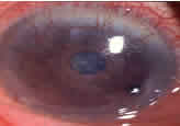
corneal ulcer
-
-
Unlike corneal erosions, in which the epithelium on the surface of the cornea is eroded, corneal ulcers occur when the corneal substance behind the epithelium is also affected, becoming cloudy or thin.
Symptoms include pain, redness, and vision loss. It is a very serious corneal disease that can lead to blindness and must be treated immediately.
corneal vascularization
-
-
This is a disease in which blood vessels advance into the cornea, which is normally vascular-free and colorless. This is a sign of severe oxygen deprivation from the eye and requires stopping contact lens wear or shortening of the wearing time.
In severe cases, there is a risk of blindness and even after treatment, the blood vessels may remain. When choosing contact lenses, select ones with good oxygen permeability to reduce the lack of oxygen in the eye. Since there are no subjective symptoms, regular eye examinations are recommended.
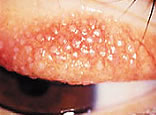
giant papillary conjunctivitis
-
-
Allergic conjunctivitis caused by lens contamination. Large bumps appear on the underside of the upper eyelid. Symptoms include itching and the tendency for contact lenses to shift easily.
It is necessary to change to lenses that are less prone to contamination, as well as to use anti-allergic medication and other eye drops and to take good care of the lenses.
corneal infiltration
-
-
It is caused by a lack of oxygen to the cornea or by inflammation of wounds caused by rubbing contact lenses.
Normally, the cornea has no blood vessels, but inflammation of the cornea causes white blood cells to collect, resulting in a cloudy white color. It is difficult to be aware of the onset of this disease because subjective symptoms are not easily recognized. Treatment involves discontinuing contact lens wear and the use of antibacterial medication.
※Many of the diseases that can occur as a result of contact problems have no subjective symptoms. Even if treatment is started, vision may not return to the best. Keratitis that can lead to blindness, may occur. Regular eye examinations are recommended.
4.Payments
-
-
After all examinations and consultations are completed, you will pay your bill at the reception counter and receive a prescription if it needed.
You can also make your next appointment at that time.
Please make an appointment if the doctor needs to follow up or if you would like to have examinations, such as a prescription for contacts or glasses. Appointments will allow for a smooth examination and consultation. You can also make an appointment by phone or online, but we recommend that you make an appointment as soon as possible at the reception counter, as appointments can fill up quickly.
If your schedule changes, please feel free to call us to reschedule your appointment.
5.Go to a pharmacy
-

-
Please bring the prescription to a pharmacy and buy the medicine.
Nearby dispensing pharmacy


 Contact Lens
contents
Contact Lens
contents